Doppler examination - Color Doppler
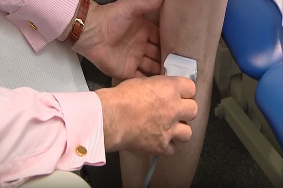
Doppler ultrasound examination is a non-invasive (painless) examination of the vascular system - both arterial and venous. This test is performed to detect abnormalities in arterial or venous blood flow caused by venous insufficiency, thrombosis in the superficial or deep system, anomalies in the structure of vessels, atherosclerosis in the arterial system, aneurysms.
Ultrasound of varicose veins
Thanks to ultrasound examination using the Doppler effect, it is possible to detect even minor diseases of arteries and veins. This examination is currently the first and necessary stage in the effective treatment of vascular diseases. It precedes the treatment of venous system diseases, both with surgical and non-operative methods. Doppler ultrasound with color flow coding is also the basic methodology for diagnosing stenosis of the carotid arteries and lower limb abdominal aortic aneurysms. Doppler ultrasound is a non-invasive test.
Color Doppler ultrasound – mechanism of action
Each ultrasound device is equipped with a transmitter, receiver, memory module and monitor. The transmitter function is performed by a special head connected with a cable to the ultrasound device. The head sends ultrasonic waves, the waves are reflected to the moving medium (flowing blood) and return to the head. The monitor records changes in the frequency of ultrasound waves reflected from flowing blood. The doctor performing the examination observes changes in the intensity and direction of blood flow on the monitor. The Doppler effect involves a change in the frequency of the sound wave received by the observer as a result of the mutual movement of the sound source and the observer. If the observer is located on an axis perpendicular to the axis of motion of the sound source (90-degree angle), the actual and perceived frequency of the sound wave are the same and the Doppler effect does not occur. In turn, the maximum Doppler effect occurs when the observer is located on the axis of motion of the sound source. Dual imaging Doppler ultrasound is based on a combination of ultrasound imaging and pulsed Doppler examination. This allows direct visualization of anatomical structures because the Doppler wave is focused on the examined vessel. During the examination, the device also performs a spectral analysis of the reflected signal, and in the case of devices with a color coding option, it also marks the flow of the blood stream with bidirectional colors (spectral analysis contains a graphic image of all the frequencies included in the Doppler signal).
Use of ultrasound
Diagnosis of blood vessels by ultrasound is performed using Doppler ultrasound. In this way, the cervical and vertebral, venous and arterial vessels of the lower limbs are checked. If the blood encounters obstacles while flowing through the veins and aorta, Doppler ultrasound will find them and allow for their quick analysis. Thanks to this, it is possible to assess the composition and shape of the atherosclerotic plaque, vascular parameters, assess the width of the arteries and the continuity of their walls, and analyze the direction of blood flow in the vessels, which should run towards the head. Duplex ultrasound technique combines a cross-sectional image with the Doppler examination result. The doctor sees the vein in which he is measuring blood flow on one half of the monitor, and the flow curve on the other half of the monitor. In this way, blood flow in each vein and artery of the lower limb can be precisely determined. In this way, all veins can be examined, superficial and deep. Thanks to this test, the doctor can determine the exact flow pattern in all veins of the limb, which is not possible using any of the other tests.
Ultrasonography of the arteries of the lower limbs is a relatively simple, non-invasive examination that allows the visualization of most pathologies and the assessment of the effectiveness of surgical treatment and postoperative complications. It also allows the lesions to be differentiated from other diseases causing symptoms of ischemia, such as arterial embolism and thrombosis, thromboembolic arteritis, and arteritis of various etiologies. In addition, ultrasound diagnostics of arteries in the lower limbs includes the assessment of the degree of dilatation of the vessel lumen, true and pseudoaneurysms, post-traumatic lesions, monitoring of surgically created fistulas and the condition of vessels during and after surgery.
What does the test look like:
A complete modern ultrasound examination of vessels consists of three basic stages, the scope of which depends on the changes detected in the vessels. The first part of the examination includes an anatomical assessment of the examined area, visualization of larger vascular trunks and determination of their topography, structure and the presence of pathological changes, including: atherosclerotic plaques, widening or narrowing. The second part of the examination, performed in a color-coded Doppler system, visualizes the flow in the selected vessel or presents a map of the vessels in the area to be diagnosed. If it is difficult to visualize the flow in the examined vessels or if there is a suspicion of the presence of small pathological vessels or the appearance of symptoms of congestion, it is necessary to perform an additional power Doppler examination. The third part of the examination consists in obtaining a record of the Doppler spectrum curve from a selected place in the vessel and making all the necessary flow measurements.
An ultrasound device is necessary to perform endovascular laser surgery. Color-marked ultrasound-guided marking of the course of ineffective venous segments (including marking the opening of the great saphenous vein to the femoral access site) undergoing the procedure is necessary to perform a safe operation. Without an ultrasound device, it is impossible to perform this procedure.
For such a diagnosis, it is necessary to have a very high-class ultrasound device, specialized for intravascular ultrasound. For this purpose, special miniature high-frequency heads are used, introduced through a catheter into the vessel lumen. They allow you to obtain an image of a cross-section of an artery, allowing you to visualize individual layers of the vessel wall and perform a detailed analysis to assess changes within it.
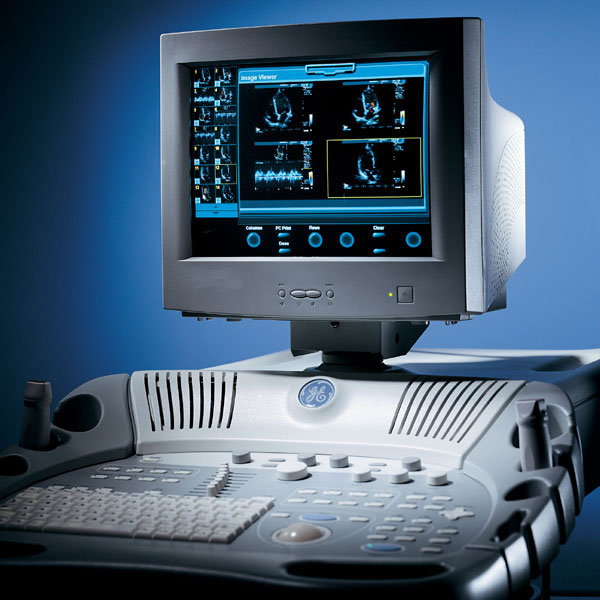
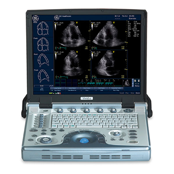
In our center, ultrasound examination is performed using the GE VIVIDe device - specialized in intravascular ultrasound and the GE VIVID3 device. The GE VIVIDe device uncompromisingly combines the highest quality of imaging, analysis and measurements with the features of an excellent intraoperative device. The system is based on the software and architecture of top-class echocardiographs - GE VIVID7, retaining all the features of archiving and post-processing, and additionally providing wireless connectivity for data transmission in computer networks. It has the ability to image and measure using the anatomical M-mode.
Because accurate diagnostics is the starting point for effective treatment, it is so important to perform an ultrasound examination of the venous system using good medical equipment and by a well-trained phlebologist.